Electroporation biopsy - QuickField simulation example
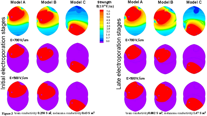
The performed numerical simulations allow to get an initial understanding of tissue areas from which e-biopsy could potentially collect the intracellular molecular markers released by electroporation. If the electric field strength thresholds for the tissues are known, the developed simulations could indicate the fractions of tumors that are electropermeabilized.
How to calculate the effect of needle-electrode position on the electric-field distribution during electroporation biopsy?
Answer Typical applications Geometry
Given
Task
Results
Joule heating of the brain during the application of pulsed electric fields for e-biopsy
Engineering question
Solve DC-conduction models for the different needle positions and compare the resulting electric-field distributions in the brain and tumor tissue.
biopsy electroporation electrodes, medical needle electrode arrays, electrochemotherapy probes
Download
Simulation problem
Problem Type
Plane-parallel multiphysics problem of DC Conduction coupled to Heat Transfer.
Brain electrical conductivity 0.258 S/m, melanoma electrical conductivity 0.43 S/m.
Reversible and irreversible electroporation thresholds for the brain are 500 V/cm and 700 V/cm respectively.
E-biopsy was performed using a combination of high-voltage short pulses 1000 V @ 40 μs with low-voltage long pulses 50 V @ 15 ms.
Pulses frequency is 4 Hz, number of pulses is 40.
Electric and thermal properties of the mice healthy brain tissue and melanoma tissue used in numerical simulations you can find here: https://doi.org/10.1371/journal.pone.0265866.s001
Study the distributions of the electric fields in the brain tissue at different positions of the needle electrode.
Electric field distribution:

Related examples